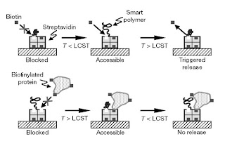
Streptavidin is a dalton protein cleansed from one of the well-known bacteria, Streptomyces avidinii. Its homo-tetramers possess an extra-high attraction for the nutrient called biotin, which is more commonly called Vitamin B7. Having a common dissociation constant, the binding process in biotin into Streptavidin substance has been acknowledged as one of the most stable non-covalent forms of interactions known to man. Streptavidin is utilized extensively in the fields of molecular biology, as well as bio-nanotechnology. This is because of the resistance of the biotin and streptavidin complex to a number of organic solvents; some denaturants like guanidinium chloride; detergents, like SDS and Triton; proteolytic enzymes; and even extreme conditions in pH and temperature.
The issue on crystallized formation of streptavidin, when it was merged with biotin was initially solved in the year 1989 through the works of Hendrickson. Consequently, solution was further developed in May 2009, with more than 134 structures placed care of Protein Databank. The C and N terminals of the full-length residue protein were processed to provide a short core streptavidin, which usually comprised of residues from 13 up to 139. On the other hand, the deletion of the termini is needed to attain high-level binding affinity of biotin. The second formation of streptavidin-type monomer is made up of 8 antiparallel beta-strands, which actually fold to provide an anti-parallel tertiary structure of its beta barrel.
A binding-site of biotin is found at an end of every β-barrel. Identical streptavidin-type monomers, like 4 identical beta-barrels are associated to provide tetra-quaternary structure of streptavidin. The binding-site located in every barrel is composed of residues that come from the barrel’s interior area. Together with the residue is the conserved Trp120 that comes from nearby sub-unit. In such process, every sub-unit contributes mainly to the binding location of the subunit. Hence the tetramer may likewise be considered as that ultimate dimer of all functional forms of dimers.
One of the streptavidin’s most common uses is the detection and purification of a wide variety of biomolecules. The stable biotin and streptavidin bond may be utilized to attach a variety of biomolecules among each other or within a stable support. Harshest forms of elemental conditions are necessary in breaking the biotin and streptavidin interaction; such process usually denatures the structure of protein meant to be purified.
Still, it was discovered that short-duration incubation with the use of water that has a temperative of above 70 degrees Celsius will break any interaction without streptavidin being denatured. This allows the re-using of solid support to streptavidin. Further steps on application are known to be Strep-tag, which is actually an optimal procedure that involves the detection and purification of certain proteins. Likewise, Streptavidin is used in conjugated blotting, as well as efficient immunoassays to a number of molecule, like horseradish peroxidase.
Targetted Immunotherapy utilizes streptavidin when conjugated to clonal antibodies against cancerous cell-specific antigen bodies. This is followed by injections of radioactive biotin, meant to deliver such radiation with full focus to cancerous cells. Hurdles involve biotin binding site saturation on Streptavidin that possesses endogenous type of biotin, and not the radioactive biotin. It also gives off a high level of exposure that is focused at the kidneys; this is because of the strong cell adsorption properties of streptavidin. Currently, it is believed that such high binding level to specific cell forms, like activated melanomas and platelets, is caused by integrin-binding happening via streptavidin RYD sequence.

No comments:
Post a Comment